0123-00 Deluxe Eight-Part Ear
0123-00 Deluxe Eight-Part Ear
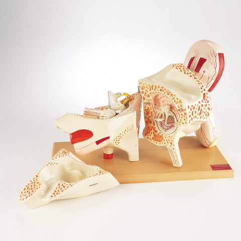
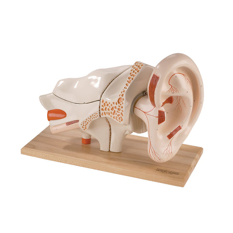
Five times life-size in unbreakable vinyl, our most detailed ear model provides a complete perspective of the middle and inner ear structures within the temporal bone.
All the skin has been removed from the detachable pinna to expose the cartilaginous structure, muscles, auricular arteries and nerves, as well as the entrance to the auditory canal.
Sections of the petrous and mastoid portions of the temporal bone can be lifted off to expose the middle and inner ear chambers.
The tympanic ring (featuring a see-through tympanic membrane) and auditory ossicles lift out of the middle chamber for close examination.
The labyrinth is extractable and dissectible into three sections. Half of it, consisting of the semicircular canals and vestibule, demonstrates the sense of balance. The semicircular canals are depicted via clear tubing filled with fluid containing a bubble that moves as the piece is tilted to demonstrate gravitational affect on the system of balance. The other half depicts the vestibular and cochlear branches of cranial nerve VIII as well as the cochlea. The cochlear part also dissects to reveal the cochlear, vestibular, and tympanic ducts.
95 hand-numbered features are identified in the corresponding key.
Overall dimensions: 17x11x8 inches (43x28x20cm)
A. External Ear
-
Auricle
-
Helix
-
Scaphoid fossa (fossa of helix)
-
Crus of helix
-
Antihelix
-
Crura of antihelix
-
Triangular fossa (fossa of
antihelix)
-
Tragus
-
Supratragic tubercle
-
Intertragic incisure
-
Antitragus
-
Anterior incisure (anterior
notch)
-
Posterior auricular sulcus
-
Cymba
-
Cavum
-
Lobule
-
Greater muscle of helix
-
Smaller muscle of helix
-
Muscle of tragus
-
Muscle of antitragus
-
Oblique muscle
-
Transverse muscle
-
Rami of anterior auricular a.
-
Rami of posterior auricular a.
-
Posterior branch of great
auricular nerve
-
-
External Acoustic Meatus
-
Cartilaginous portion
-
Osseous portion
B.
Middle Ear
III. Tympanic Membrane withTympanic Ring
-
Anterior portion of membrane
-
Posterior portion of membrane
-
Pars flaccida
-
Anterior malleolar ligament
-
Posterior malleolar ligament
-
Radiating fibers of
membranous layer
-
Circular fibers of membranous
layer
IV. Tympanic Cavity 34. Malleus
a. Head
b. Neck
c. Manubrium35. Incus
d. Bodye. Short crus
f. Long crus 36. Stapes
g. Anterior crus h. Posterior crus i. Base
-
Tympanic antrum
-
Entrance of Eustachian
(auditory) tube
-
Facial nerve, cut across
-
Tensor tympani muscle
-
Promontory
-
Round (cochlear) window
-
Oval (vestibular) window
-
Mastoid air cells
C. Inner Ear
V. Semicircular canals45. Cavity of semicircular canals 46. Cavity of cochlea
47. Internal auditory meatus 48. Anterior semicircular canal 49. Posterior semicircular canal 50. Lateral semicircular canal 51. Round (cochlear) window 52. Oval (vestibular) window 53. Ampulla of anterior canal 54. Ampulla of posterior canal 55. Ampulla of lateral canal56. Common crus 57. Utriculus
58. Sacculus
59. Vestibular nerveVI. Cochlea
-
Beginning of first turn of
cochlea
-
Second turn of cochlea
-
Cupula of cochlea
-
Vestibular lip
-
Tympanic lip
-
Spiral lamina
-
Cochlear nerve to branch of au-
ditory nerve (Vestibulocochlear
nerve-Cranial Nerve VIII)
-
Vestibular nerve
-
Cochlear nerve
-
Facial nerve (Cranial Nerve VII)
-
Geniculate ganglion
VII. Structures of the Temporal Bone 71. Mastoid process
72. Eustachian (auditory) tube 73. Styloid process74. Glenoid fossa
75. Internal carotid artery
76. Groove for sigmoid sulcus 77. Mastoid portion of temporalbone
78. Petrous portion of temporalbone
-
-
-
We Also Recommend